2026 Top Arthroscopic Surgery Equipment Types?
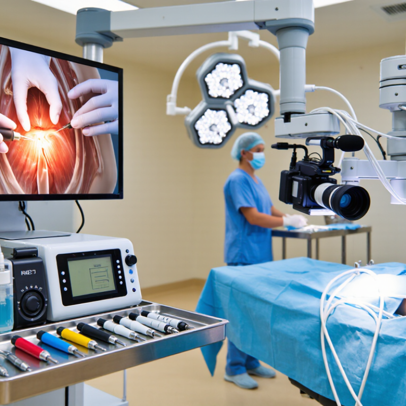
Arthroscopic surgery equipment is becoming more specialized, connected, and procedure-focused as orthopedic techniques advance in 2026. This guide examines the main equipment types used in modern arthroscopy, from high-definition cameras and light sources to fluid management systems, shavers, radiofrequency tools, and surgical pumps. Each category supports a different part of the procedure. Together, these systems help clinicians view, access, repair, and protect joint structures through small incisions.
Practical selection requires more than comparing prices or product images. Surgeons often assess image clarity, instrument control, irrigation stability, ergonomic design, and compatibility with existing towers. Nurses and biomedical engineers also evaluate setup time, cleaning requirements, maintenance access, and staff training. A bright monitor is useful. Stable fluid flow matters more than expected. Reliable performance can reduce interruptions during delicate procedures, especially when visibility changes around bleeding tissue or narrow joint spaces.
However, no system is perfect. A feature-rich device may increase complexity, training demands, or service costs. A compact tool may improve handling but limit certain procedures. Product claims should be reviewed against clinical evidence, manufacturer documentation, user experience, and applicable safety standards. The most suitable arthroscopic surgery equipment depends on the joint treated, surgical technique, facility resources, and team expertise. The categories also overlap, and real operating-room decisions are rarely as simple as a product chart suggests. Careful evaluation remains essential.
Arthroscopic Surgery Equipment: Definition and Clinical Purpose
2026 Top Arthroscopic Surgery Equipment Types?
Arthroscopic surgery equipment supports minimally invasive joint diagnosis and treatment. An arthroscope uses a small camera, light source, and lens system to display the joint on a monitor. Surgeons may add fluid pumps, shavers, radiofrequency instruments, graspers, and motorized burrs. Each tool has a distinct clinical purpose.
The equipment must create a clear, stable, and controlled surgical field. A pump maintains joint distension, while the camera reveals cartilage, ligaments, and synovial tissue through a small portal. Shavers remove damaged tissue. Graspers retrieve loose fragments. Energy instruments can manage selected soft-tissue bleeding, but their settings require careful judgment.
Grand View Research reported that the global arthroscopy devices market was valued at approximately USD 6 billion in 2023, with continued growth expected through 2030. This expansion reflects rising sports injuries, aging populations, and demand for shorter hospital stays. Market forecasts are useful, though they do not prove better patient outcomes. That distinction matters.
Clinical experience shows that equipment selection depends on anatomy, procedure type, surgeon skill, and facility resources. A sophisticated tower cannot correct poor visualization or weak training. The system also requires sterilization, maintenance, and compatible accessories. Small failures matter. A fogged lens can disrupt an entire procedure. Future designs may become smarter, but human review remains essential.
Core Components of a Modern Arthroscopy System
2026 Top Arthroscopic Surgery Equipment Types?
Core Components of a Modern Arthroscopy System
A modern arthroscopy system begins with the arthroscope, a narrow camera inserted through a small portal. Its lens, viewing angle, and image resolution affect tissue recognition during surgery. A camera control unit converts the optical signal into a clear image for the surgical monitor. Accurate color matters. Blood, cartilage, and synovial tissue should remain visually distinct under changing light conditions.
The light source must provide stable illumination without excessive heat near the scope tip. Fluid management equipment controls irrigation pressure and keeps the joint cavity open. Pumps should respond smoothly when pressure changes, especially during shoulder or knee procedures. A shaver system removes damaged tissue, while radiofrequency instruments can address selected soft-tissue work. These devices require careful settings, trained handling, and reliable safety checks.
Small details matter. Foot pedals should be easy to identify through sterile drapes, and cables should not cross the surgeon’s working path. Recording systems support documentation, teaching, and later review, but stored images still need secure handling. Sterile cannulas, graspers, punches, and probes must match the procedure and reprocessing requirements. A bright image can still mislead when the lens is dirty or the fluid is cloudy. No setup is flawless. Teams should verify image quality, pressure readings, instrument function, and backup equipment before incision, then reflect honestly on failures after each case.
2026 Top Arthroscopic Surgery Equipment Types: Core Components of a Modern Arthroscopy System
Arthroscopy systems combine visualization, access, tissue management, irrigation, and energy-control equipment. The chart compares representative outer diameters commonly used for arthroscopes and working instruments; actual dimensions vary by joint, procedure, and manufacturer.
Top Arthroscopic Equipment Types Used in 2026
Top Arthroscopic Equipment Types Used in 2026
Arthroscopic surgery in 2026 relies on several connected equipment types. High-definition arthroscopes provide the viewing path through small portals. Camera heads, light sources, and surgical monitors convert that view into usable detail. Fluid management pumps maintain joint expansion and help clear blood or debris. Shaver systems remove damaged tissue with controlled suction. Radiofrequency devices support soft-tissue treatment, but their heat requires careful monitoring.
Grand View Research estimated the global arthroscopy devices market at approximately US$6.5 billion in 2023. Its report projects continued growth through 2030, supported by sports injuries, aging populations, and minimally invasive procedures. Fortune Business Insights also identifies arthroscopes, implants, fluid systems, and visualization equipment as major market segments. These figures show demand, not automatic clinical value.
In operating rooms, equipment selection should match the joint, procedure, and surgeon’s experience. A stable pump can matter more than a brighter display. Sterile tubing, hand instruments, burrs, cannulas, and imaging accessories remain essential. Small details matter. Poor cable placement can disrupt workflow. Excessive irrigation pressure may affect tissue visibility or patient safety. I have seen equipment lists look impressive while missing practical backup tools. That gap deserves reflection. More technology is not always better surgery.
Specialized Tools for Different Joint Procedures
In 2026, arthroscopic equipment is becoming more procedure-specific. Knee surgery commonly requires a high-definition camera, fluid management pump, shaver, and meniscal repair instruments. Shoulder procedures need curved suture passers, anchors, and flexible cannulas for labral or rotator cuff work. Small joints demand slimmer optics and delicate graspers. Every millimeter matters.
Hip arthroscopy uses longer scopes, traction-compatible instruments, and angled burrs for femoroacetabular impingement. Ankle procedures often depend on compact scopes, small shavers, and low-profile probes. Wrist and elbow surgery need narrow access tools, because nerves and vessels leave little room for error. It looks simple on a tray. It is not.
Grand View Research reports continued growth in the arthroscopy instruments market, supported by minimally invasive treatment demand and improved visualization systems. Fortune Business Insights also projects steady expansion in the global arthroscopy devices market through 2032. These reports support investment, but market forecasts are not surgical evidence. Clinical teams should match tool diameter, viewing angle, irrigation control, and tissue protection to each joint. I have seen equipment lists become excessive. More instruments do not automatically create better surgery. Training, sterilization reliability, and fast access during a procedure may matter more than adding another specialized device.
How to Compare Arthroscopic Equipment for Clinical Use
When comparing arthroscopic equipment for clinical use, start with the procedure, not the catalogue. A 4 mm rigid scope may suit large joints, while smaller optics improve access in narrow spaces. Check image resolution, field of view, and fog resistance under real operating-room lights. A sharp image matters when cartilage edges look pale and wet. Test the camera, light source, and monitor as one system. Compatibility is often overlooked.
For tissue management, compare shavers by blade control, suction stability, vibration, and cleaning access. A fluid pump should maintain a clear field without creating excessive joint pressure. Energy devices need precise activation and dependable insulation checks. Ask how quickly staff can change settings during a case. Foot pedals should be easy to identify through sterile drapes. Small interface errors become large delays.
Procurement teams should review sterilization instructions, service response, training, and total cost per procedure. Reusable instruments may reduce waste, yet they demand strict inspection and documented reprocessing. Disposable components can simplify turnover, but their cost deserves careful modeling. Run a hands-on trial with surgeons, nurses, and technicians. A comparison checklist may change after observing cable damage or awkward tray layouts. That is an uncomfortable lesson. Performance claims still need local evidence, not confident wording.