>

Top 10 Hip Arthroscopy Procedures for Global Buyers

Hip arthroscopy procedures are moving from specialist operating rooms into a broader global healthcare market. Buyers now compare more than price. They assess visualization, instrument reliability, surgeon training, after-sales service, and regulatory documentation. A smooth procedure may depend on details as small as a clear 70-degree scope or stable fluid pressure.

Industry reports show steady expansion. Grand View Research identifies sports injuries, improved imaging, and minimally invasive treatment as key market drivers. Fortune Business Insights also forecasts continued growth in the global hip arthroscopy market. Their estimates differ, however. The figures are useful, but not perfectly aligned. Buyers should question market projections before making large purchasing decisions.

Hip preservation specialist Thomas L. Byrd, MD, has described this evolution clearly: “Hip arthroscopy has evolved from a diagnostic procedure to a therapeutic procedure.” That shift explains the growing demand for labral repair systems, femoroacetabular impingement instruments, cartilage treatment tools, and loose-body removal equipment. The International Society for Hip Arthroscopy emphasizes structured training, careful patient selection, and consistent surgical technique. Those points matter across borders.

This guide reviews the top 10 hip arthroscopy procedures for global buyers. It connects clinical purpose with practical purchasing needs. Product catalogs can look impressive. Real-world performance may tell a different story. Buyers should examine published evidence, sterilization requirements, surgeon feedback, and local regulatory approval before choosing a supplier. Availability is not the same as clinical value.
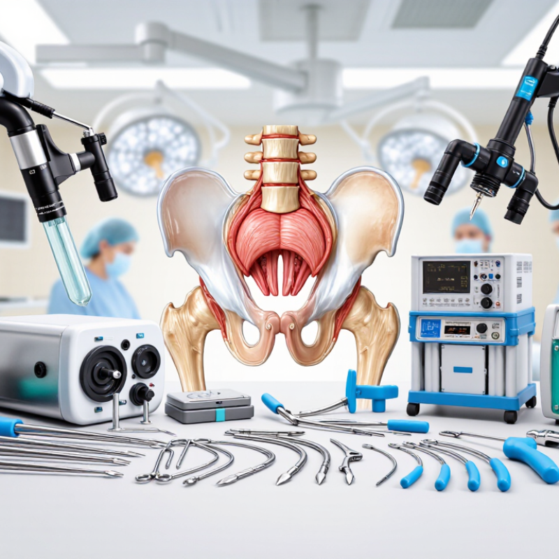
Top 10 Hip Arthroscopy Procedures for Global Buyers

Hip Arthroscopy: Purpose, Scope, and Global Buyer Considerations

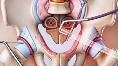
Hip arthroscopy is a minimally invasive procedure used to inspect and treat problems inside and around the hip joint. Its scope commonly includes ten procedures: diagnostic arthroscopy, labral repair, labral debridement, cam osteoplasty, pincer osteoplasty, loose-body removal, synovectomy, cartilage treatment, ligamentum teres treatment, and capsular closure or plication. Each option serves a different clinical purpose. Surgeons select procedures according to imaging, symptoms, joint stability, cartilage condition, and patient activity.

Global buyers should evaluate systems through clinical workflow, not catalog size. Instruments must support clear visualization, controlled tissue handling, reliable access, and efficient operating-room turnover. Cannulas, graspers, shavers, burrs, suture tools, pumps, and traction accessories should work as a coordinated set. Staff training also matters. A technically advanced device may underperform without proper positioning, sterile processing, and hands-on instruction.

Fit matters. Hospitals should review surgeon experience, local repair patterns, maintenance support, sterilization requirements, and regulatory documentation before purchasing. Compatibility with existing towers and operating-room equipment can reduce unnecessary costs. Buyers should request performance data, training records, warranty terms, and post-market safety information. Evidence varies. Some procedures depend heavily on patient selection and surgeon judgment, so outcome claims deserve careful review. No checklist fits every hospital. A lower-cost option may create delays if instruments are fragile, incomplete, or difficult to reprocess. Procurement teams should compare total ownership costs and real clinical needs, not only the initial quotation.
Top 10 Hip Arthroscopy Procedures for Global Buyers

Hip Arthroscopy: Purpose, Scope, and Global Buyer Considerations

The chart compares commonly performed hip arthroscopy procedures using representative operating-room planning times in minutes. Actual duration varies according to pathology, surgical technique, patient anatomy, equipment, and surgeon experience. Buyers should evaluate instrument compatibility, implant requirements, reusability, sterilization workflow, and local regulatory requirements before procurement.

Core Diagnostic and Access Procedures in Hip Arthroscopy

Hip arthroscopy begins with accurate diagnosis and controlled access. Surgeons commonly inspect the central compartment, peripheral compartment, labrum, cartilage, ligamentum teres, and synovium. Diagnostic arthroscopy, capsular access, traction-assisted distraction, and portal placement form the technical foundation for these procedures. A 2023 systematic review in Arthroscopy analyzed more than 6,000 hip arthroscopies and linked structured examination with better identification of labral and cartilage injuries.

The anterolateral portal usually provides the viewing route, while the mid-anterior and distal anterolateral portals support instruments. Fluoroscopy can confirm needle position and reduce unintended joint penetration. The Danish Hip Arthroscopy Registry has reported meaningful improvements in patient-reported pain and function after properly selected procedures, although outcomes vary with cartilage damage and arthritis. Small details matter, especially when the joint space is narrow.

For global buyers, core procedure sets should support diagnostic viewing, safe traction, portal dilation, capsular management, synovectomy, loose-body removal, labral repair, acetabular rim work, femoral osteoplasty, and microfracture. These are not interchangeable steps. A 2022 review in the Journal of Hip Preservation Surgery noted that patient selection strongly influences reported success rates. That remains easy to underestimate. Access can look routine on paper, yet a misplaced portal may restrict visualization or irritate nearby nerves. Experienced teams still review imaging, traction time, instrument reach, and surgeon training before procurement.

Top 10 Hip Arthroscopy Procedures for Global Buyers - Core Diagnostic and Access Procedures in Hip Arthroscopy
No. Procedure Primary Clinical Purpose Typical Indications Common Surgical Access or Portal Use Core Instruments and Consumables Typical Case Complexity Key Procurement Considerations
1 Diagnostic Hip Arthroscopy Direct visualization and systematic assessment of the central and peripheral compartments of the hip joint. Unexplained hip painSuspected labral injuryCartilage damage Usually begins through anterolateral and modified anterior portals under traction; additional portals may be established as required. Arthroscope, camera system, light source, inflow tubing, cannulas, guidewires, spinal needle, shaver, and fluid management accessories. Basic to intermediate Clear optics, reliable fluid control, compatible cannula sizes, and sterile tubing availability are important for consistent visualization.
2 Central Compartment Access Provides controlled entry to the acetabulum, femoral head, labrum, and articular cartilage under traction. Labral pathologyChondral lesionsLoose bodies Commonly uses an anterolateral viewing portal and an anterior working portal; portal placement is confirmed with imaging and direct visualization. Traction table or traction device, portal needles, guidewires, dilators, cannulas, obturators, and arthroscopic pump tubing. Basic to intermediate Portal systems should support controlled dilation, secure soft-tissue protection, and compatibility with standard hip arthroscopy instruments.
3 Peripheral Compartment Access Allows treatment of structures outside the central joint without or after reducing traction. Cam morphologyCapsular pathologySynovitis Access is typically performed through established lateral or anterior portals with the hip flexed, depending on the target anatomy. Flexible or standard cannulas, blunt obturators, radiofrequency probes, shavers, burrs, and fluid-management components. Intermediate Equipment should allow stable access in a flexed hip and provide adequate soft-tissue protection around the femoral neck.
4 Acetabular Labral Repair Reattaches and stabilizes a torn labrum to restore the suction seal and support hip joint function. Detached labrumFemoracetabular impingementTraumatic tears Usually performed through anterior and anterolateral working portals after evaluating the labrum and acetabular rim. Suture anchors, drill guides, flexible or rigid suture passers, suture management instruments, graspers, and knot-management tools. Intermediate to advanced Anchor size range, insertion accuracy, pullout performance, suture handling, and availability of nonabsorbable sutures are key purchasing factors.
5 Labral Debridement Removes unstable or irreparable labral tissue while preserving as much viable tissue as possible. FrayingPartial-thickness tearsDegenerative tissue Performed through anterior or anterolateral portals with direct visualization of the acetabular rim and labral surface. Small-joint shaver blades, arthroscopic punches, graspers, radiofrequency probes, suction tubing, and cannulas. Basic to intermediate Shaver blade control, low-profile instruments, tissue-preserving suction, and dependable fluid evacuation help reduce iatrogenic damage.
6 Femoroplasty Reshapes an abnormal femoral head-neck junction to reduce cam-type impingement during hip motion. Cam morphologyLoss of head-neck offsetImpingement-related labral injury Usually performed in the peripheral compartment through anterolateral and anterior portals, often with fluoroscopic or arthroscopic assessment. High-speed burr, motorized handpiece, protective cannula, flexible probe, fluoroscopy-compatible positioning equipment, and irrigation tubing. Intermediate to advanced Burr durability, thermal control, visibility of bone removal, handpiece reliability, and compatibility with imaging workflows are important.
7 Acetabuloplasty Resects excessive acetabular rim bone to correct pincer-type impingement while protecting the labrum and joint surface. Pincer morphologyAcetabular overcoverageRim prominence Typically uses anterior and anterolateral portals to expose the acetabular rim and perform controlled bone trimming. Arthroscopic burr, motorized handpiece, bone-cutting accessories, radiofrequency probe, suture anchors when labral repair is required, and irrigation equipment. Intermediate to advanced Precise burr control, low-profile access, reliable irrigation, and instruments that support incremental rim resection are essential.
8 Loose Body Removal Retrieves free fragments from the hip joint to relieve mechanical symptoms and prevent further cartilage damage. Osteochondral fragmentsSynovial chondromatosisPost-traumatic fragments Access may involve central or peripheral compartment portals; additional portals can be created when fragments are difficult to reach. Large-bore cannulas, graspers, retrieval baskets, flexible probes, shavers, suction tubing, and fluid-management accessories. Basic to advanced, depending on fragment size and location Portal diameter options, grasper strength, retrieval capacity, and reliable visualization in a distended joint are important.
9 Chondroplasty Stabilizes unstable cartilage flaps and removes damaged cartilage that may cause mechanical irritation. Focal chondral defectsCartilage flapsEarly degenerative changes Performed through central compartment portals after systematic inspection of the acetabular and femoral head cartilage. Small-diameter shaver, arthroscopic curette, probe, graspers, radiofrequency device when appropriate, and continuous irrigation. Basic to intermediate Fine instrument control, low suction settings, atraumatic probes, and clear visualization are needed to preserve stable cartilage.
10 Synovectomy Removes inflamed or hypertrophic synovial tissue and improves visualization of the joint surfaces. SynovitisInflammatory arthropathyPigmented synovial disease May be performed in both central and peripheral compartments through standard portals, with additional access based on disease distribution. Motorized shaver, radiofrequency probe, graspers, biopsy forceps when tissue sampling is indicated, cannulas, and fluid-management tubing. Intermediate Shaver efficiency, bleeding control, tissue containment, suction performance, and availability of biopsy-compatible instruments support procedural consistency.
Note: Procedure selection, portal configuration, traction use, imaging support, and required instruments vary according to patient anatomy, pathology, surgeon training, and institutional protocols.

Labral Repair, Reconstruction, and Related Treatments

Top 10 Hip Arthroscopy Procedures for Global Buyers

Labral repair is often selected when the hip labrum is torn but still suitable for preservation. Surgeons use small portals, a camera, and fine instruments to reattach the labrum near the acetabular rim. The goal is to restore the seal around the joint and reduce painful catching. Debridement may remove unstable tissue, but it can be less protective than repair in selected patients. This distinction deserves careful discussion.

Labral reconstruction uses a graft when tissue loss, severe fraying, or previous surgery limits repair. Related procedures may include acetabuloplasty, femoroplasty, capsular closure, cartilage treatment, loose-body removal, synovectomy, and psoas-related treatment. These procedures address different causes of impingement, instability, or irritation. Combining treatments can be appropriate, but more treatment is not automatically better. A detailed examination and imaging review remain essential.

Global buyers should request the surgeon’s qualifications, procedure-specific experience, implant or graft details, and rehabilitation plan. Ask how complications, follow-up imaging, and unexpected findings will be managed across borders. Recovery may involve crutches, restricted hip motion, and months of strengthening. Timelines vary widely. Published success rates can also look clearer than real-world outcomes. Patient age, cartilage condition, bone shape, muscle control, and prior operations all influence results. A technically successful repair may still disappoint if expectations are unrealistic.

Impingement, Cartilage, and Loose-Body Procedures

Top 10 Hip Arthroscopy Procedures for Global Buyers

Hip arthroscopy addresses several causes of pain through small portals around the joint. Common procedures include diagnostic arthroscopy, cam osteoplasty, pincer acetabuloplasty, and combined impingement correction. Surgeons may also perform labral repair, labral reconstruction, cartilage debridement, chondroplasty, microfracture, and loose-body removal.

Impingement procedures reshape excess bone that restricts hip movement. Cam correction treats a bony prominence on the femoral head-neck junction. Pincer correction removes excessive acetabular coverage. Accurate imaging matters here. A few millimeters can affect surgical planning. Labral repair restores the seal around the socket, while reconstruction may help when tissue quality is poor. Results depend on patient selection, technique, and rehabilitation.

Cartilage procedures require careful judgment. Debridement can remove unstable fragments, while chondroplasty smooths damaged surfaces. Microfracture creates small openings that may encourage fibrocartilage formation. It is not equivalent to restoring normal cartilage. Loose-body removal extracts free fragments that cause catching, locking, or sudden pain. Buyers should review instrument compatibility, sterilization methods, training support, and regulatory documentation. Experienced teams also assess imaging quality and postoperative protocols. No device replaces clinical judgment. A practical concern remains: purchasing a technically advanced system without adequate staff training may increase delays, errors, or inconsistent outcomes.

How Global Buyers Compare, Source, and Select Hip Procedures

Global buyers comparing hip arthroscopy procedures need more than a ranked list. Common options include labral repair, femoroacetabular impingement correction, cartilage treatment, loose-body removal, synovectomy, and ligamentum teres procedures. Each option serves a different clinical finding. A reliable comparison starts with imaging protocols, diagnosis criteria, expected recovery, and published outcome data. Buyers should ask whether the proposed procedure matches patient selection, not just equipment availability. That question often exposes weak sourcing decisions.

During sourcing, procurement teams can request surgeon training records, facility accreditation, sterilization controls, traceable instruments, and complication reporting. They should compare total care pathways, including anesthesia, rehabilitation, follow-up imaging, and revision planning. A low procedure price may hide travel, translation, or extended recovery costs. Clear regulatory documentation matters across borders. So does communication. Ask for evidence in plain language. Then verify it independently.

Selection should use a weighted scorecard, but the scorecard is never perfect. Clinical fit deserves more weight than marketing claims. Buyers can review infection rates, unplanned admissions, return-to-activity timelines, and patient-reported outcomes. They should also examine how complications are managed after patients return home. Procurement reviews often reveal gaps that brochures avoid. Follow-up ownership may remain unclear. Price still matters, but it should not lead the decision. A careful buyer leaves room for uncertainty and revises the shortlist when new evidence appears.