>

How to Compare Hip Arthroscopy Procedures in 2026?

Choosing among hip arthroscopy procedures in 2026 requires more than comparing incision size or recovery promises. It requires careful attention to diagnosis, surgical technique, surgeon experience, rehabilitation, and patient goals. The AAOS Appropriate Use Criteria for femoroacetabular impingement emphasizes matching treatment decisions with symptoms, imaging, physical findings, and prior care. That framework matters because a painful hip does not always have one clear cause.

Professional reports also reveal a complicated picture. The International Society for Hip Preservation Surgery consensus work supports structured assessment for labral injuries, impingement, and cartilage damage. Meanwhile, systematic reviews published in Arthroscopy report meaningful improvements in pain and hip-function scores after selected procedures. However, outcomes vary widely. Age, arthritis, bone shape, athletic demands, and rehabilitation access can change the result. A market report may show rising demand, but demand alone cannot prove clinical value.

Marc J. Philippon, MD, a leading hip-preservation surgeon, famously said, “The hip is the new frontier in sports medicine.” His observation captures the field’s momentum, but it also invites caution. New tools are not automatically better tools. In this guide, we compare common hip arthroscopy procedures through clinical purpose, evidence quality, recovery timelines, risks, and long-term expectations. Small details matter, such as whether imaging shows cartilage loss or whether a patient can follow a six-week protected rehabilitation plan. No comparison is perfect. Some evidence remains uneven, and patients should discuss uncertain findings with a qualified orthopedic specialist before choosing treatment.

How to Compare Hip Arthroscopy Procedures in 2026?

What Hip Arthroscopy Treats and When Comparison Is Appropriate

How to Compare Hip Arthroscopy Procedures in 2026?

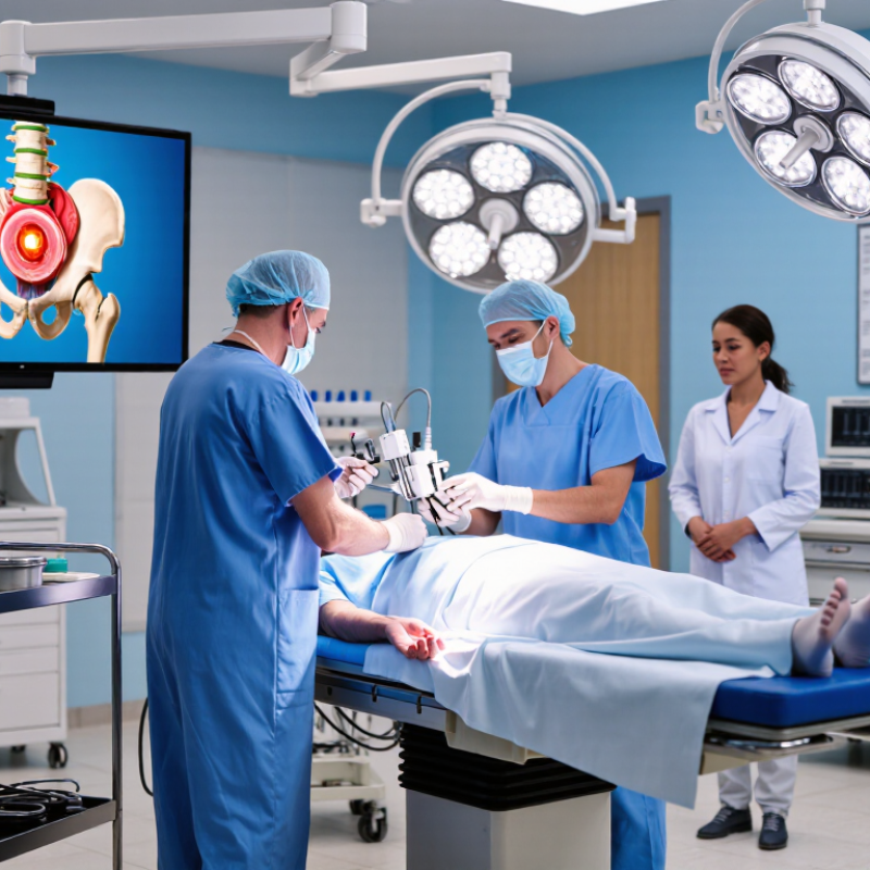
Hip arthroscopy uses small instruments to treat problems inside the hip joint. Common conditions include labral tears, femoroacetabular impingement, loose bodies, and selected cartilage injuries. The procedure may repair or reshape damaged tissue, depending on the cause. Details matter.

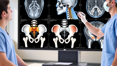
Comparison becomes appropriate only after a careful diagnosis. A specialist should review symptoms, physical findings, X-rays, and sometimes magnetic resonance imaging. Two procedures may sound similar but address different problems. A labral repair aims to preserve tissue, while labral reconstruction may be considered when the tissue cannot be repaired. These choices depend on damage, age, activity, and previous treatment.

Do not compare procedures by name alone. Ask how each option addresses the confirmed problem, expected pain relief, mobility, and rehabilitation time. Surgical experience matters, but so does honest discussion of alternatives, including structured physiotherapy. Recovery is not perfectly predictable. Some patients improve steadily, while others need longer rehabilitation. I would not treat a scan alone as a decision. Examination findings and daily limitations must also guide the plan. No comparison is perfect. A reliable consultation should explain risks, possible complications, and what happens if the first treatment does not provide enough improvement.

How to Compare Hip Arthroscopy Procedures in 2026?

What hip arthroscopy treats and when comparison is appropriate

How to read this comparison

The chart shows commonly used ranges for protected or restricted weight-bearing after selected hip arthroscopy procedures. Labral repair and femoroacetabular impingement correction often require approximately 2–4 weeks, while microfracture usually requires a longer period to protect the treated cartilage.

These are typical clinical ranges, not universal rules. The appropriate procedure depends on the diagnosis, cartilage condition, hip stability, symptoms, imaging findings, athletic goals, and the surgeon’s rehabilitation protocol.

Labral Repair

Treats a repairable acetabular labral tear and aims to restore the suction-seal function of the labrum.

FAI Correction

Reshapes excess bone associated with cam or pincer-type femoroacetabular impingement.

Microfracture

Addresses selected small full-thickness cartilage defects by stimulating a healing response.

Ligamentum Teres Reconstruction

May be considered for selected cases of persistent instability or symptomatic ligamentum teres deficiency.

Clinical ranges vary according to the procedure performed, tissue quality, concurrent repairs, patient factors, and postoperative assessment.

Key Differences Among Hip Arthroscopy Techniques

Comparing hip arthroscopy techniques requires more than counting portal incisions. The main differences involve labral treatment, cartilage management, femoral reshaping, and capsular handling. Labral repair preserves the sealing function around the hip joint. Debridement removes unstable tissue but may sacrifice healthy structure. A 2024 systematic review in Arthroscopy reported lower revision and total hip replacement rates after repair than after debridement, although much of the evidence remained observational.

Femoroacetabular impingement surgery usually combines cam or pincer correction with labral treatment. The amount of bone removed matters. Too little correction may leave painful contact; too much can weaken stability. Capsular closure may also reduce postoperative instability, especially in patients with lax joints. However, technique selection should follow imaging, cartilage condition, hip stability, and the surgeon’s measured experience, not a fashionable procedure name. The UK FASHIoN randomized trial, published in BMJ, found a 10.6-point greater improvement in iHOT-33 scores after arthroscopy than physiotherapy at 12 months, among 348 participants. That result supports surgery for selected patients, but it does not prove one arthroscopic technique suits everyone.

In practical terms, ask how the surgeon protects the labrum, controls bone removal, and measures recovery. Look for published patient-reported outcomes. Also ask about complications and revision rates. Evidence is improving, but follow-up periods remain uneven. Some comparisons still look clearer than they really are.

How to Compare Hip Arthroscopy Procedures in 2026? - Key Differences Among Hip Arthroscopy Techniques

Typical clinical ranges and rehabilitation milestones are general estimates for uncomplicated cases; actual treatment depends on imaging, cartilage status, surgeon preference, and patient factors.
Technique Primary target Common indications Core surgical steps Typical loading and activity progression Key advantages Important limitations or risks
Labral repair Acetabular labrum Traumatic or degenerative labral tears, especially when the labral tissue is repairable and the hip has treatable structural abnormalities. The torn labrum is mobilized, the acetabular rim is prepared, and the labrum is secured with suture anchors. Associated impingement or instability may be addressed during the same operation. Protected weight bearing commonly lasts about 2–4 weeks, with range-of-motion restrictions during early healing. Strengthening progresses over approximately 6–12 weeks; return to pivoting sport often takes about 4–6 months. Preserves the suction-seal function of the labrum and is generally preferred over removing viable labral tissue. Healing may be compromised by severe cartilage damage, poor tissue quality, untreated dysplasia, residual impingement, or excessive postoperative loading.
Labral debridement Unstable or nonviable portions of the labrum Small, frayed, or irreparable tears, particularly when the remaining labral tissue is stable and the joint has limited degenerative change. Loose or damaged tissue is trimmed and the stable labral margin is preserved. Concomitant bony impingement may be corrected if present. Weight bearing is often advanced more quickly than after repair, sometimes as tolerated, although restrictions depend on additional procedures. Low-impact exercise may begin within several weeks; higher-impact activity commonly requires 2–4 months. Usually involves a simpler repair construct and may allow faster early rehabilitation. Removing too much tissue can reduce the suction seal and may be less suitable for large tears, instability, or a viable repairable labrum.
Femoroplasty Femoral head–neck junction Cam-type femoroacetabular impingement, in which an aspherical femoral-head junction contacts the acetabular rim during hip motion. The prominence is reshaped under arthroscopic visualization, usually with dynamic assessment to reduce impingement while avoiding excessive bone removal. Partial weight bearing may be used for about 2–4 weeks, especially when combined with labral repair. Running often resumes around 3–4 months, with sport-specific return commonly around 4–6 months. Addresses the mechanical source of impingement and can improve hip flexion and rotation when patient selection is appropriate. Under-resection can leave residual impingement; over-resection may weaken the femoral neck or contribute to instability. Advanced arthritis generally reduces expected benefit.
Acetabuloplasty Acetabular rim Pincer-type femoroacetabular impingement or focal acetabular overcoverage confirmed by clinical examination and imaging. Excess rim bone is carefully resected, often followed by labral repair. The amount of resection is planned to avoid creating undercoverage or instability. Protected weight bearing commonly lasts about 2–4 weeks; strengthening usually advances over 6–12 weeks, while unrestricted sport may require 4–6 months. Can reduce abnormal rim contact and restore space for hip flexion when overcoverage is focal and accurately identified. Excessive resection may cause instability or dysplasia-related symptoms. Global acetabular deficiency may require a different reconstructive approach rather than isolated arthroscopy.
Labral reconstruction Absent, severely damaged, or irreparable labrum Irreparable labral deficiency, failed prior surgery, or substantial loss of the native suction seal in a hip without advanced arthritis. A graft is shaped and fixed around the acetabular rim to recreate the labral seal. Any residual bony impingement or instability must also be assessed and treated. Rehabilitation is commonly more protective than isolated debridement; partial weight bearing may last about 3–6 weeks, with return to unrestricted sport often around 5–8 months. Provides a reconstructive option when native tissue cannot reliably be repaired and aims to restore the suction seal. Technically demanding, with graft-related considerations, longer rehabilitation, and less uniform long-term evidence than primary repair.
Microfracture Focal full-thickness cartilage defect Small, contained chondral defects without diffuse end-stage osteoarthritis, often treated together with impingement correction or labral surgery. The unstable cartilage is removed to create stable edges, and small perforations are made in the underlying bone to release marrow elements. Limited or non-weight bearing commonly lasts about 4–8 weeks, depending on defect size and location. Impact activities may be delayed for approximately 4–6 months. Can stimulate formation of repair tissue without requiring a graft or implanted cartilage construct. The repair tissue is fibrocartilage rather than normal hyaline cartilage; results are less predictable for large lesions, diffuse arthritis, or high-impact athletes.
Capsular closure or plication Hip joint capsule Capsular laxity, microinstability, connective-tissue laxity, or situations in which a larger capsulotomy could increase instability risk. The capsulotomy is closed, and redundant capsule may be tightened with sutures. The amount of tightening is individualized to preserve necessary motion. Protected weight bearing and avoidance of extension and external rotation are common during the first 2–4 weeks. Progressive strengthening may take 8–12 weeks or longer. May preserve or improve passive stability and is particularly relevant when instability is a concern. Over-tightening can restrict motion and contribute to stiffness; failure to identify underlying dysplasia or structural deficiency can lead to persistent symptoms.
Comparison note: Hip arthroscopy commonly combines more than one technique—for example, labral repair with femoroplasty or acetabuloplasty. Procedure selection should be based on symptoms, physical examination, radiographs, magnetic resonance imaging, cartilage condition, hip coverage, and the presence of arthritis or instability.

How Surgeons Evaluate Risks, Benefits, and Patient Eligibility

Comparing hip arthroscopy procedures in 2026 requires more than matching an MRI finding to a surgical technique. Surgeons assess the patient’s pain pattern, walking mechanics, hip motion, strength, and daily limitations. A visible labral tear may not explain every symptom. Sometimes it is an incidental finding.

Eligibility depends on the suspected cause and the joint’s overall condition. For femoroacetabular impingement, a surgeon may consider reshaping abnormal bone and repairing damaged tissue. A labral repair may preserve function better than removing unstable tissue, but the choice depends on tissue quality, cartilage health, and activity goals. Significant arthritis can reduce the likely benefit of arthroscopy. Age alone should not decide eligibility. It still matters.

Risk assessment includes infection, blood clots, nerve irritation, traction-related discomfort, persistent pain, and possible revision surgery. Surgeons also review previous treatments, medical conditions, medications, and rehabilitation commitment. A careful consultation should explain expected recovery, restrictions, and realistic alternatives, including physiotherapy or observation. Ask how the diagnosis was confirmed. Ask what happens if symptoms continue.

No scorecard is perfect. Even experienced surgeons can face unexpected cartilage damage during surgery. That uncertainty deserves honest discussion. In practice, the strongest decision combines examination findings, appropriate imaging, surgical experience, and the patient’s own priorities. A technically successful procedure may still disappoint someone seeking immediate return to sport. Recovery is rarely linear.

Comparing Recovery Timelines, Rehabilitation, and Expected Outcomes

How to Compare Hip Arthroscopy Procedures in 2026?

A useful comparison starts with the tissue being treated. Labral repair often allows gradual weight-bearing, while femoroacetabular impingement correction may require similar protection but longer strength work. Cartilage procedures can follow stricter restrictions. The operation name alone does not predict recovery.

The early phase usually focuses on pain control, gentle range of motion, and safe walking. Many patients use crutches for several days or weeks, depending on surgical findings. Physical therapy then targets hip stability, core control, and gait quality. Progress should be measured by movement and function, not time alone. Slow progress happens.

A recreational athlete may return to low-impact exercise within a few months, while cutting, jumping, or competition can take longer. Desk work may resume earlier, but prolonged sitting can still irritate the hip. Expected outcomes depend on age, cartilage condition, muscle strength, and adherence to rehabilitation. Evidence supports careful patient selection, yet outcome studies do not perfectly match every individual. That matters.

Before comparing procedures, ask how much bone or cartilage will be treated, what restrictions are expected, and which milestones guide advancement. A qualified hip specialist should explain risks, alternatives, and the evidence behind the recommendation. Be cautious with promises of a fixed return date. Recovery is rarely linear. Pain can improve before strength returns, creating false confidence. Regular reassessment can identify stiffness, weakness, or persistent symptoms before they become harder to address.

How to Assess Costs, Surgeon Expertise, and Long-Term Value

Comparing hip arthroscopy in 2026 requires more than reviewing the advertised surgical fee. Request a written estimate covering consultation, imaging, anesthesia, facility charges, implants, medication, and physical therapy. Check what insurance may exclude. A low initial quote can become expensive after extra appointments or delayed recovery. Ask about payment policies and possible revision costs. Clarity matters.

Surgeon expertise should be assessed with evidence, not confidence alone. Review training, certification, annual procedure volume, and experience with your specific problem. Ask how often patients develop infection, stiffness, nerve symptoms, or persistent pain. Request realistic recovery timelines, including when walking, driving, work, and sports may resume. A surgeon who explains uncertainty may be more trustworthy than one promising perfect results. Short answers deserve questions.

Long-term value includes function, comfort, and the chance of avoiding another procedure. Compare published outcomes with your age, activity level, cartilage condition, and diagnosis. Ask whether non-surgical care has been fully considered. Physical therapy can affect both results and total cost. I would not judge value from price alone. That shortcut is tempting, but incomplete. Keep copies of imaging reports, consent documents, and follow-up plans. Discuss the decision with a qualified clinician who can examine you directly, because online comparisons cannot reveal every risk. Recovery is not always linear. Reassess the plan if pain, weakness, or mobility changes unexpectedly.